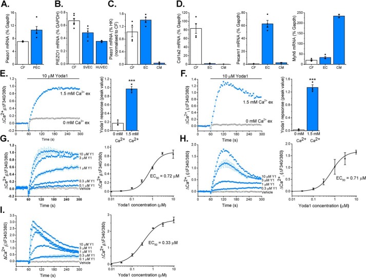
Figure 1.
Piezo1 is expressed by cardiac fibroblasts and forms a functional ion channel. A and B, RT-PCR analysis of Piezo1 mRNA expression in murine cardiac fibroblasts (CF, n = 3) compared with murine pulmonary endothelial cells (PEC, n = 5) (A) and human CF (n = 6) compared with human saphenous vein endothelial cells (SVEC, n = 3) and HUVECs (n = 3) (B). Expression was measured as percent of the housekeeping control (Gapdh/GAPDH). C, RT-PCR analysis of Piezo1 mRNA expression in fibroblast-enriched fraction 2 (CF) and endothelial cell–enriched fraction 1 (EC) isolated from murine heart using the MACS technique (n = 4). Cardiomyocytes (CM) were isolated from separate hearts (n = 2). Expression was measured relative to three housekeeping genes (Gapdh, Actb, and Hprt) and normalized to the CF sample. D, RT-PCR analysis of cell type-specific marker genes for CF (Col1a2), endothelial cells (Pecam1), and cardiomyocytes (Myh6) in MACS fractions as for C. E and F, representative Ca2+ traces and mean ± S.E. are shown. Ca2+ entry was evoked by 10 μm Yoda1 in murine (E) and human (F) cardiac fibroblasts in the presence or absence of extracellular Ca2+. ***, p < 0.001 (paired t test; n/N = 3/9). G–I, Ca2+ entry evoked by varying concentrations of Yoda1 application at 60 s, ranging from 0.1–10 μm in murine (G) and human (H) cardiac fibroblasts and HEK T-Rex-293 cells heterologously expressing mouse Piezo1 (I). Vehicle control is illustrated by the black trace. Mean ± S.E. are displayed as concentration–response curves, and fitted curves are plotted using a Hill equation, indicating the EC50 of Yoda1 (n/N = 3/9).

