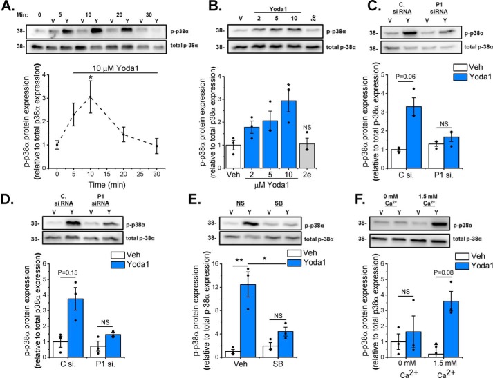
Figure 7.
Yoda1-induced p38 MAPK phosphorylation depends on Piezo1. Cellular protein samples were immunoblotted for p-p38α and reprobed with antibody for total p38α to confirm equal protein loading. The bar charts show mean densitometric data of p-p38α normalized to p38α expression. A, murine cardiac fibroblasts (n = 6) treated with DMSO vehicle (V) or 10 μm Yoda1 (Y) for 5–30 min. *, p < 0.05 versus vehicle-treated cells (repeated measures one-way ANOVA, p = 0.0085). B, murine cardiac fibroblasts (n = 3) treated for 10 min with varying concentrations of Yoda1 (2–10 μm) or 10 μm compound 2e. *, p < 0.05; NS, not significant versus vehicle-treated cells (repeated measures one-way ANOVA, p = 0.0122). C and D, murine (C) or human (D) cardiac fibroblasts (n = 3) transfected with either scrambled or Piezo1-specific siRNA before treatment with vehicle or 10 μm Yoda1 for 10 min. Repeated measures two-way ANOVA for C: p = 0.0202, F = 48.0 (Yoda1); p = 0.3045, F = 1.87 (siRNA); p = 0.0903, F = 9.6 (interaction). Repeated measures two-way ANOVA for D: p = 0.0282, F = 34.0 (Yoda1); p = 0.2669, F = 2.32 (siRNA); p = 0.478, F = 0.75 (interaction). Post hoc test: not significant. E, murine cardiac fibroblasts (n = 3) exposed to 10 μm SB203580 for 1 h before treatment with vehicle or 10 μm Yoda1 for 10 min. Repeated measures two-way ANOVA: p = 0.0312, F = 30.6 (Yoda1); p = 0.5898, F = 0.40 (SB203580); p = 0.0148, F = 65.9 (interaction). Post hoc test: **, p < 0.01; *, p < 0.05. F, murine cardiac fibroblasts (n = 3) treated for 10 min with vehicle or 10 μm Yoda1 in either standard DMEM or DMEM containing 1.75 mm EGTA to chelate free Ca2+. Repeated measures two-way ANOVA: p = 0.0123, F = 79.5 (Yoda1); p = 0.5739, F = 0.44 (Ca2+); p = 0.128, F = 6.34 (interaction). Post hoc test: not significant.

