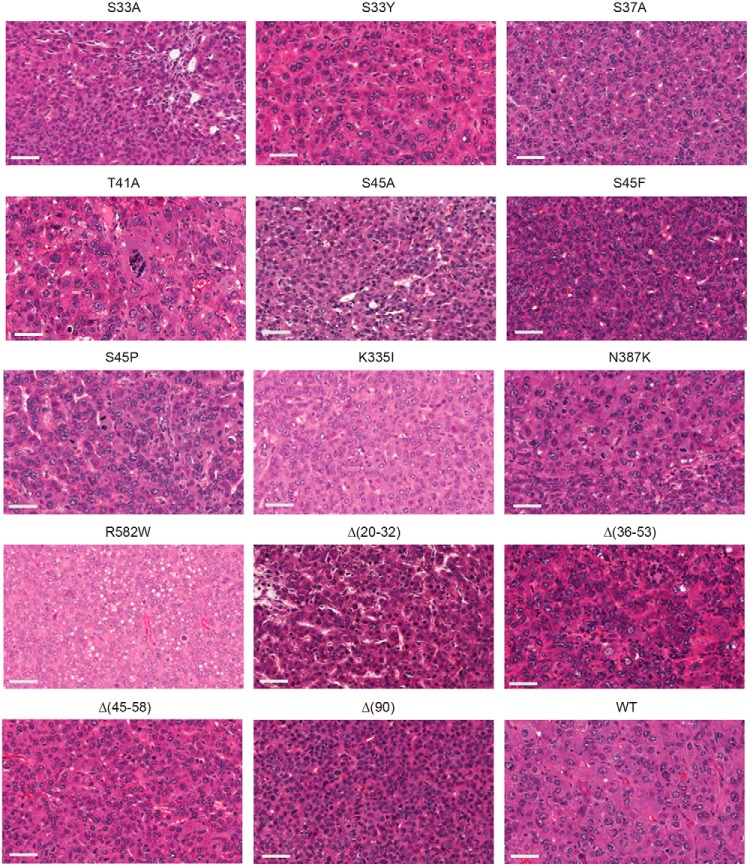
Figure 2.
Histology of β-catenin mutant tumors. At least three tumors induced by each β-catenin mutant were examined under blinded conditions. Typical hematoxylin and eosin–stained sections are depicted here. Bars, 50 μm. The most prominent HB histologic subtypes observed in each tumor cohort are summarized in Table 1.

