Figure 4.
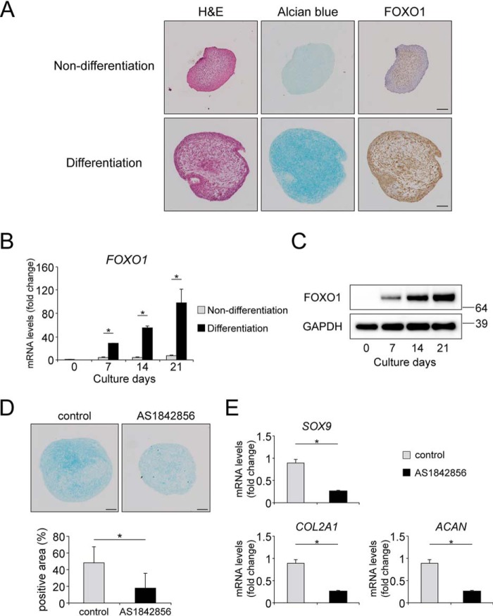
FOXO1 regulates TGFβ1-induced chondrogenic differentiation in hMSCs. Pellets of hMSCs were incubated for 21 days in normal medium not including TGFβ1 (Non-differentiation) or chondrogenic medium including TGFβ1 (differentiation). A, representative images of: left, hematoxylin and eosin (H&E) staining; center, Alcian blue staining; and right, FOXO1 on day 21. Bars represent 100 μm. B, relative mRNA levels of FOXO1 were measured by qRT-PCR. Gene expression at each stage is given relative to the level on day 0; n = 3. C, time course of expression of FOXO1 protein in pellets incubated in chondrogenic medium for 21 days, as determined by Western blotting. D, Alcian blue staining images of pellets differentiated in chondrogenic medium with or without AS1842856 (0.1 μm) for 21 days. The graph below shows the percentages of Alcian blue-positive areas. Bars represent 100 μm; n = 6. E, pellets of hMSCs were differentiated in chondrogenic medium with or without AS1842856 (0.1 μm) for 21 days. Relative mRNA levels of SOX9, COL2A1, and ACAN were measured by qRT-PCR. Gene expression is given relative to the level in cultures incubated without AS1842856; n = 4. Data are presented as mean ± S.D. Statistical analysis was performed using Wilcoxon's rank-sum test. *, p < 0.05.

