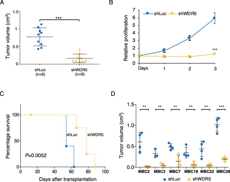
Fig. 1.
WDR5 controls tumorigenesis in breast cancer. a MCF10DCIS cell line was infected to silence WDR5 (shWDR5) or a neutral control (shLuc) and inoculated in vivo in immunocompromised mice. When tumors of control group reached maximum volume expected, all the mice were sacrificed to compare effect on tumor growth. Dot plots represent tumor volume in shWDR5 or shLuc MCF10DCIS (n = 9 per group; mean ± SD—cm3). Statistical significance was determined using an unpaired Student t test (***P < 0.001). b In vitro relative proliferation values of shWDR5 in MCF10DCIS cells are reported with respect to control (shLuc). Statistical significance on n = 3 experiments was determined using an unpaired Student t test (***P < 0.001). c Percentage of survival of mice belonging to control or shWDR5 groups was calculated. Mice were sacrificed when each tumor reached maximum volume expected. Differences among groups were calculated by using Log-rank test (n = 5 per group; P = 0.0052). d Six MBC PDXs were infected to target control (shLuc) and shWDR5. Transduced cells were transplanted in NSG mice. Dot plots represent volume (mean ± SD—cm3) of three to four tumors per group. Statistical significances were calculated by applying an unpaired Student t test (**P < 0.01; ***P < 0.001)

