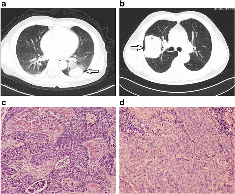
Fig. 1.

Representative CT and HE images of LELC and squamous carcinoma On CT scans, LELC usually presented a round-like well-defined mass/nodule (a); while squamous carcinoma was relatively larger with an irregular shape (b). For the histopathological examination with HE staining, a large island of nested tumor cells infiltrated by lymphocytes were found in LELC (c, ×200), whereas in squamous carcinoma, squamoid tumor cells were poorly-differentiated with abundant eosinophilic cytoplasm (d, ×200)
