Fig. 5.
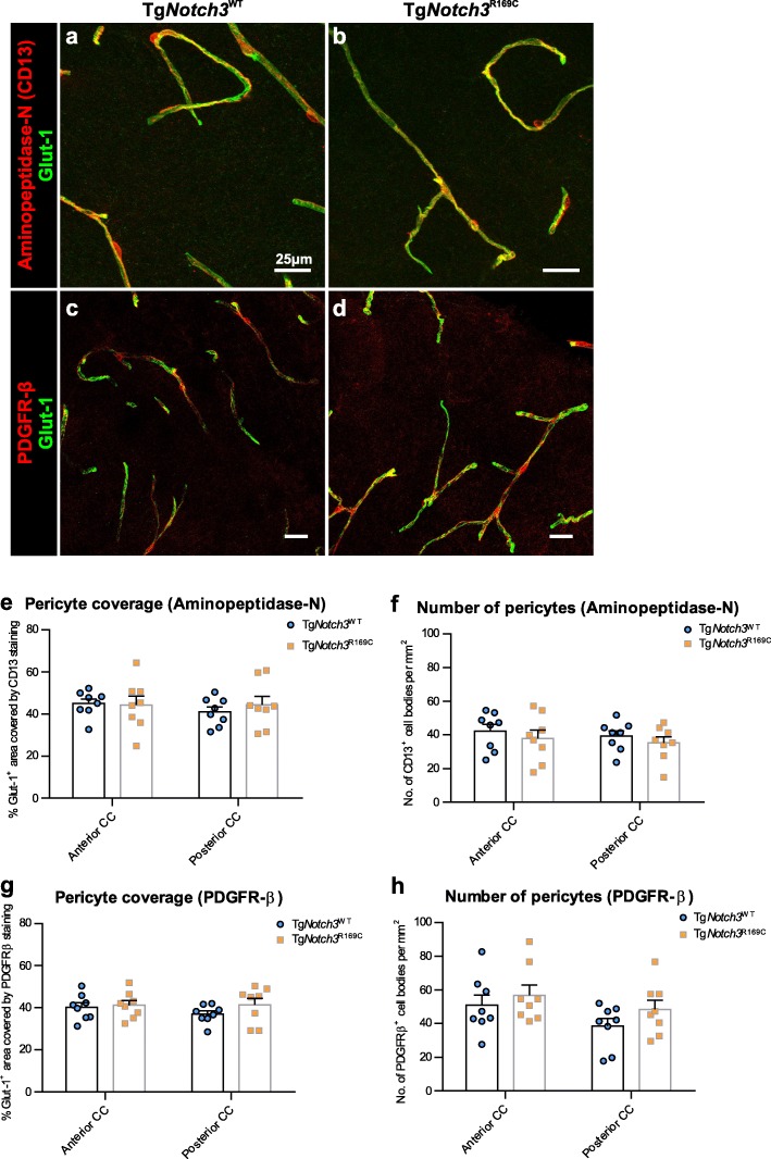
Normal pericyte coverage and number in the white matter of CADASIL mice. Immunofluorescent images showing capillaries (glut-1; green) and pericytes (a-b: aminopeptidase-N; c-d: PDGFR-β; red) in the anterior corpus callosum (CC) of TgNotch3WT (a, c) and TgNotch3R169C (b, d) mice. Quantification of pericyte coverage (e: aminopeptidase-N; g: PDGFR-β) and pericyte number (f: aminopeptidase-N; h: PDGFR-β) shows no difference in the anterior or posterior CC. Mean ± SEM; two-way ANOVA with Tukey’s post-hoc tests; n = 8. Scale bar: 25 μm

