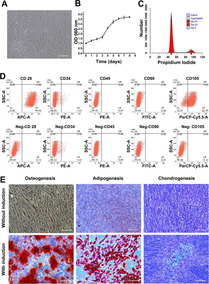
Fig. 1.
The characteristics of hASCs. a The fourth-passage hASCs screened by light microscopy (scale bar 100 μm). b The growth curve was determined by CCK-8 assays. hASCs were in the slow growth phase from the first to the third days, exponential growth phase from the third to the sixth days, and the plateau phrase from the seventh d. c hASC cell cycle analysis by flow cytometry: G1 phase (82.12% ± 5.83%) and S/G2 phase (17.88% ± 2.59%). d Immunophenotypes of hASCs detected by flow cytometry: CD29(+) (99.81% ± 0.76%), CD90(+) (98.43% ± 1.77%), CD105(+) (92.21% ± 6.48%); CD34(−) (2.39% ± 0.72%), CD45(−) (0.97% ± 0.41%). e Multilineage of hASCs induced by media. The mineralized nodules were stained with Alizarin Red. The lipid droplets were stained with Oil Red O. The proteoglycans were stained with Alcian Blue (scale bar 100 μm)

