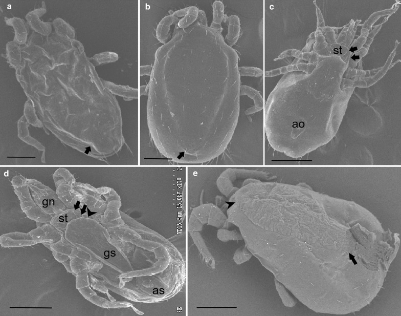
Fig. 3.
Comparative SEM of dorsal and ventral view of NFM, TFM and PRM. a Dorsal view of NFM with a posterior end tapering acutely (arrow). b The posterior end tapers more evenly in TFM (arrow). c Ventral view of NFM with the genitoventral (epigynal) shield attenuate or narrowly rounded posteriorly and a distinct sternal shield (st) with two pairs of setae (arrows). d Ventral view of the NFM with two pairs of setae on the sternal plate (arrows) and a third pair of setae on the unsclerotized integument (arrowhead). e Dorsal view of PRM, showing the idiosoma broadly rounded posteriorly (arrow) and prominent shoulder (arrowhead); adults PRM are generally larger in size than NFM and TFM. Scale-bars: a, c, e, 300 μm; b, d, 200 μm. Abbreviations: gn, gnathostoma; st, sternal plate; ao, anal opening; as, anal shield

