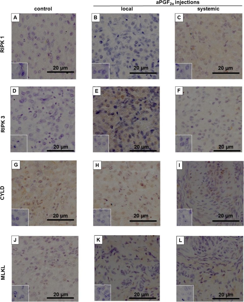
Fig. 8.
Representative section of images of localization of: (a, b, c) RIPK1, (d, e, f) RIPK3, (g, h, i) CYLD and (j, k, l) MLKL protein in the bovine early-stage corpora lutea (CL) at 4 h after local or systemic PGF2α analogue (aPGF2α) administration. Each small window shows a negative control stained with normal rabbit IgG instead of primary antibody. Positive immunohistochemistry staining was assessed as brown staining. Bar = 20 μm

