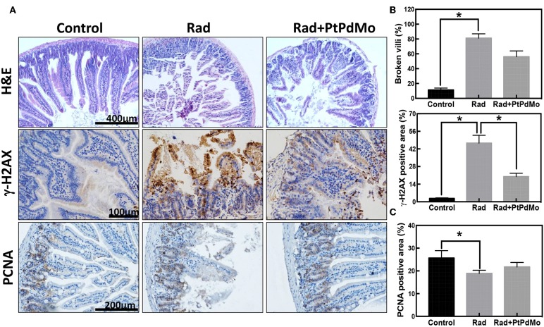
Figure 6.
(A) Pathology study carried 7 days after radiation using H&E staining. (B) Immunochemistry staining of γ-H2AX and (C) Immunochemistry study of PCNA 7 days after radiation. Positive area was calculated using IHC tool box in ImageJ software (Data was analyzed using one-way ANOVA, and presented as mean ± SD, *p < 0.05). The mice were exposed to 137Cs-γ ray with dosage of 7.2 Gy, and the dose rate was 1.0 Gy/min.

