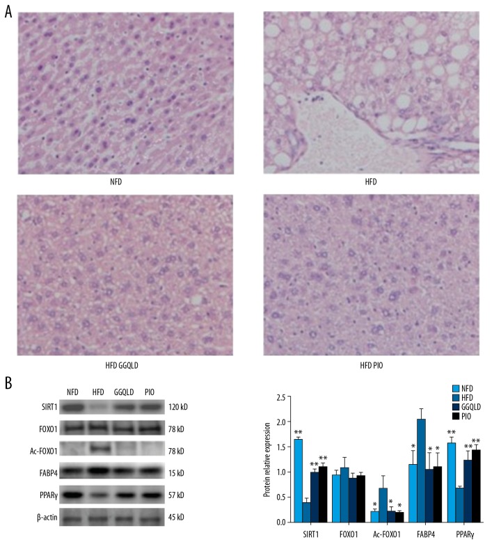
Figure 4.
GGQLD improved histopathology and affected the expression levels of key molecules in the SIRT1/FOXO1 signaling pathway in liver. (A) Liver tissues were subjected to paraffin sectioning and HE staining. For each group, liver samples from 3–5 mice were used. Representative images are shown (×200). (B) Total proteins of liver tissues were extracted and subjected to Western blot analysis for the detection of SIRT1, FOXO1, Ac-FOXO1, FABP4, and PPARγ. Representative images are shown (left). For quantification, optical densities of the bands were determined. β-actin was used as a loading control. Normalized protein expression levels were calculated. Data from 3 independent experiments were used for statistics, and results are expressed as the mean±SEM (right). * P<0.05 vs. HFD, ** P<0.01 vs. HFD.

