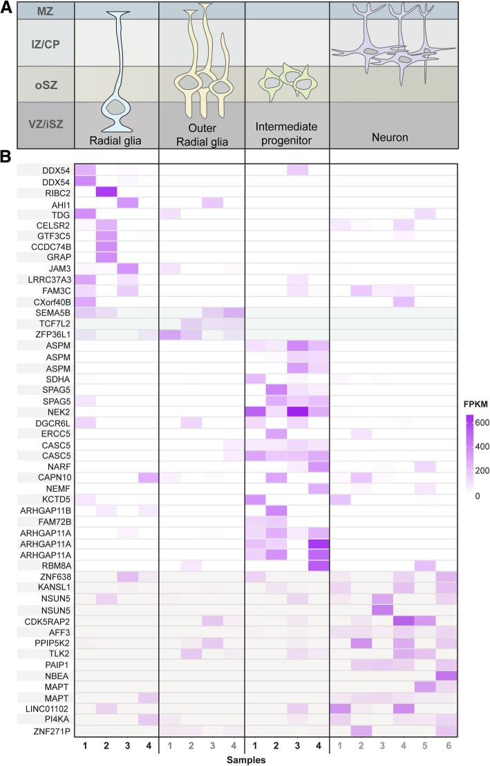
Fig. 3.
a The upper panel depicts differentiation stages of human radial glia cells. Rows represent the correspondent brain location and columns represent the different cell types along a virtual timeline. Cell location starts from the inner layer of the ventricular zone (the inner subventricular zone VZ/iSZ) and goes through the outer subventricular zone (oSZ) to the region between the intermediate zone and the cortical plate (IZ/CP). b The heatmap is a graphical representation of normalized read counts (FPKM values) across all samples for each transcript of highly expressed genes with human-specific features. Color intensity varies according to FPKM value as shown in the scale. Highly expressed genes were defined as the top 2000 in terms of FPKM and those that were highly expressed in at least one of the four cell stages were selected. The first 12 genes are highly expressed in radial glial cells, the next 3 genes (light grey shade) in outer radial glia, the further 15 in intermediate progenitor cells and the last 13 (light grey shade) in differentiated neurons. The columns of the heatmap represent different samples of each cell type (the 4 radial glia samples, 4 outer radial glia samples, 4 intermediate progenitor samples and 6 neuron samples)

