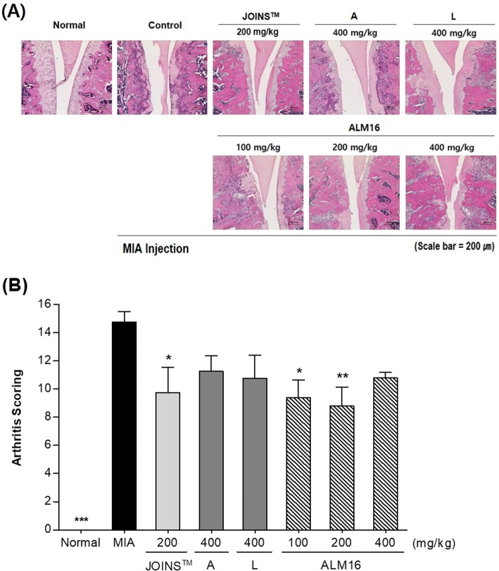
Fig. 8.
Histopathological changes of articular cartilage the femoral-tibial knee joint from MIA-induced OA rat. Wister rats were subjected to intra-articular injection of MIA (1 mg/animal) and fed orally with or without samples daily for 2 weeks. a Histological sections (× 200) were stained with hematoxylin & eosin (H&E) staining. b Histopathological changes are quantitatively expressed by arthritis scoring. The results are expressed as the mean ± S.E.M. Data were analyzed by one-way ANOVA Tukey’s test to compare all of the tested groups. *p < 0.05, **p < 0.01, ***p < 0.001 compared with the MIA-induced control group

