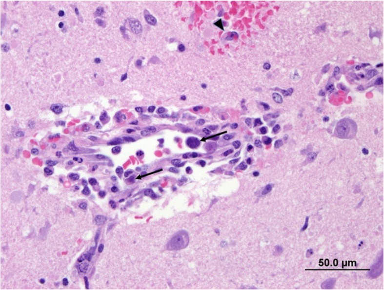
Fig. 1.

Photomicrograph of vessel in the brain. The wall is infiltrated by macrophages with perivascular accumulation of red blood cells, small amount of fibrin and macrophages. Small haemorrhage in the neuropil and erythrophagocytosis (arrowhead). Endothelial cells contain large basophilic intranuclear inclusion bodies (arrow). H & E stain. Bar = 50.0 μm
