Fig. 2.
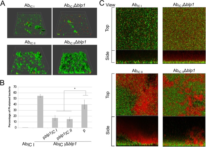
CLSM analysis of biofilms formed by the A. baumannii IC I and IC II strains. a – 3D visualization of initial attachment to the plastic by A. baumannii blp1 gene deletion mutants and parental strains assessed after 2 h of incubation; bacteria were stained with SYTO9 (green) and propidium iodide (red). b – number of propidium iodide stained bacteria after 2 h of incubation, compared to the total amount of cells and expressed as a percentage; error bars represent standard errors from the measurements of six different CLSM pictures, significance was assessed by t-test, (*P < 0.05). c – mature biofilm formation after 24 h of incubation; A. baumannii parental strains and blp1 gene deletion mutants were stained with SYTO9 (green) and propidium iodide (red); representative top and side views are given

