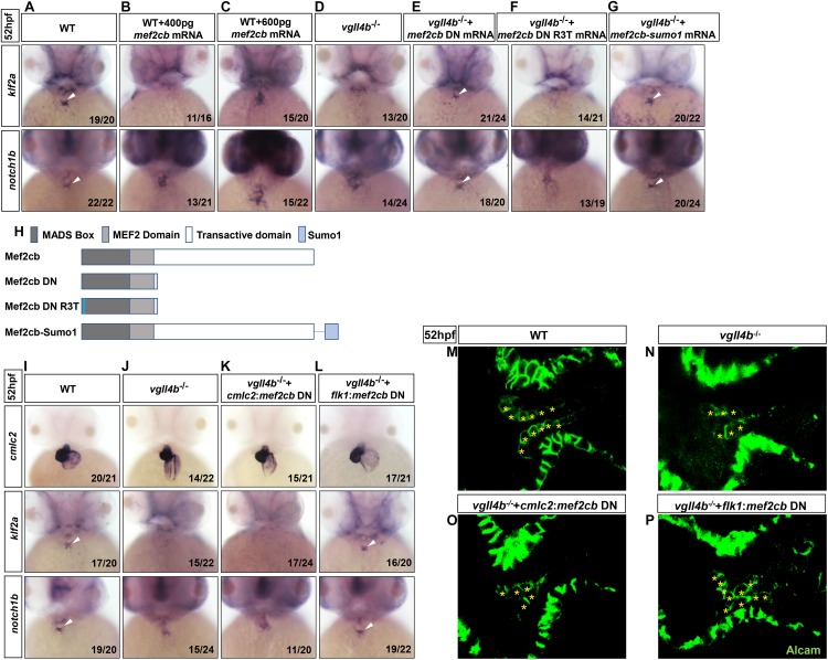
FIGURE 4.
Aberrant activation of Mef2c due to the disruption of Vgll4b-Mef2c complex, accounts for the valvulogenesis defects in vgll4b mutants. (A–C) Mef2cb mRNA was injected into one-cell stage wild type embryos. (D–G) Mef2cb DN, mef2cb DN R3T, and mef2cb-sumo1 mRNA rescue assays in vgll4b–/– embryos. (H) Schematic representation of variant forms of Mef2cb, including WT, DN, R3T, and Sumo1 fusion mutants. (I–L) Cmlc2:mef2cb DN or flk1:mef2cb DN plasmid rescue assays in vgll4b–/– embryos. (M–P) Representative images show Alcam staining of sibling and vgll4b–/– embryos rescued with cmlc2:mef2cb DN or flk1:mef2cb DN plasmid at 52 hpf. The endocardial cushion cells expressing Alcam were indicated by asterisks. Scale bar: 10 μm.

