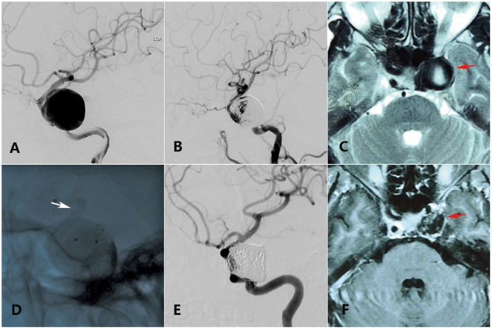
Figure 1.
A 55-year-old woman with a giant ophthalmic segment ICA aneurysm presented with visual loss and incomplete ONP of the left side showing visual improvement merely in the last clinical follow-up. (A) Preoperative DSA shows a giant ophthalmic segment ICA aneurysm. (B) Immediate postoperative DSA shows loose embolization of aneurysm. (C) Preoperative axial-plane MRI shows an ophthalmic segment ICA aneurysm (red arrow). (D) Intraoperative DSA shows placement of a PED (white arrow) covering the aneurysmal neck. (E) Thirteen-month DSA shows variation of the coils in the sac. (F) Twenty-five-month axial-plane MRI shows shrinkage of the aneurysm (red arrow).

