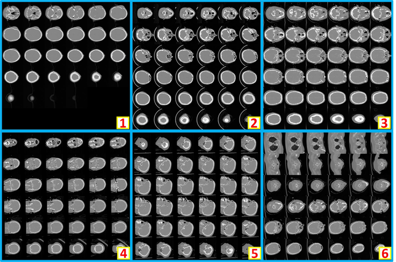
Figure 2.
The example montage images of usable whole brain 3D CT scans. “1” shows a montage image with < 36 slices after zero padding. “2” and “3” are the montage images resampled from axial view images, while “4” and “5” are from coronal and saggital view images respectively. “6” presents an example that whole brain and part of body are included in one scan. As the entire brain volume is included, “6” is also regarded as usable.

