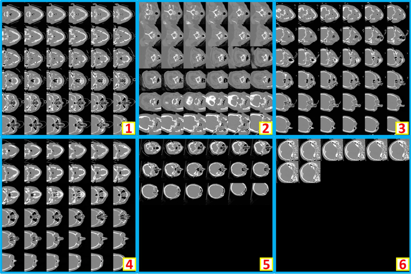
Figure 4.
This figure shows six representative examples of unusable scans. “1” to “4” shows the incomplete brains (e.g., maxillofacial CT, neck CT, temporal bone CT, patient movement). In “5”, an abnormal spatial translation is found in last two slices. For “6”, the scan only covers few slices of the cranium.

