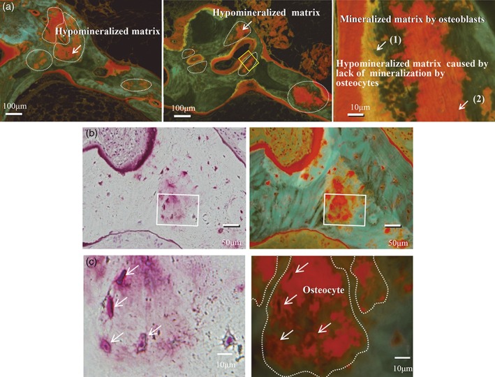
Figure 4.

(A) Hypomineralized bone area before and after PTX in group III (upper panels). Increased hypomineralized bone area 4 weeks after PTX was found in a group III patient not receiving alfacalcidol administration. The schema on the right shows both the single labelings caused by osteoblasts and hypomineralized bone area caused by osteocytes. These results indicate that vitamin D is required at least in moderate doses for adequate mineralization by osteocytes and osteoblasts. Mineralization in the osteocytic perilacunar/canalicular system was not observed around the osteocyte lacunae in a patient not receiving alfacalcidol. White arrows are pointing to (1) mineralized matrix by osteoblasts, and (2) hypomineralized matrix caused by lack of mineralization by osteocytes. (B, C) Higher‐resolution images of osteocyte lacunae. Higher‐resolution images of osteocyte lacunae within the hypomineralized bone area, as well as osteoblasts on top of the surface of the respective area, are shown. White arrows are pointing to hypomineralized matrix area.
