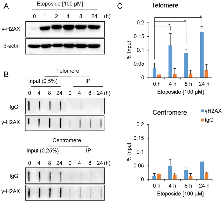
Fig 1. Induction of telomere damage upon etoposide treatment.
A. Immunoblot of γ-H2AX in HeLa cells treated with etoposide for the indicated times; β-actin was used as a loading control. B. ChIP results for telomeric and centromeric DNA with the anti-γ-H2AX antibody in HeLa cells treated with etoposide for the indicated times. DNA slot blots were hybridized with telomere- or centromere-specific probes. Antibodies specific for γ-H2AX and control IgG were used for the ChIP assays. C. Quantification of three independent ChIP assays represented in B (mean ± standard deviation, SD). Mann-Whitney U-test was used to compare differences in DNA levels between each time point and 0 h; *P < 0.05.

