
Fig 2.
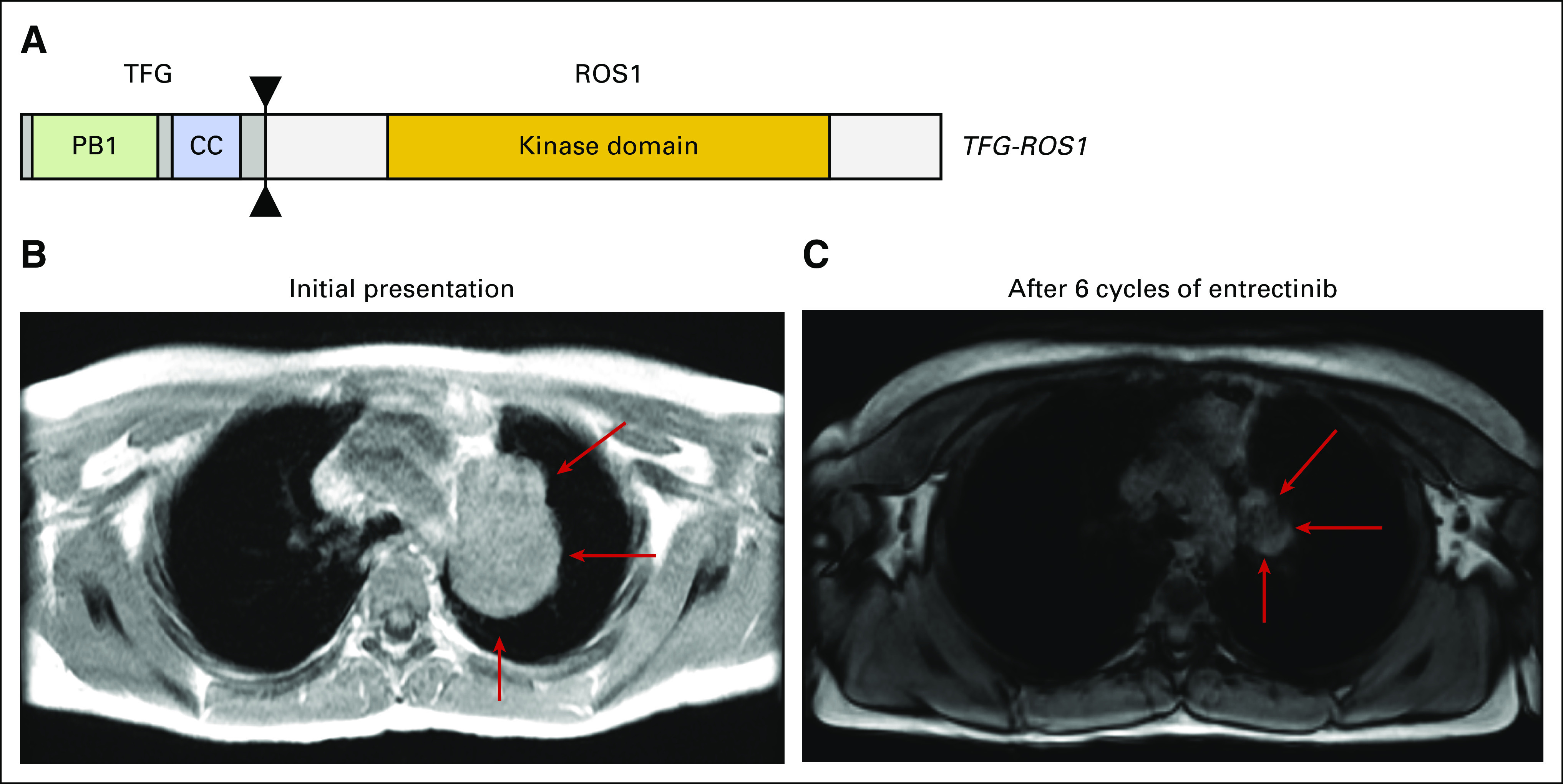
(A) TFG-ROS1 rearrangement, resulting in an in-frame fusion between TFG exon 4 and ROS1 exon 35. Contrast-enhancing magnetic resonance imaging scan of the chest of patient 2 taken from axial 3D DualEcho sequences (B) at baseline (measurements: 5.8 × 4.2 cm; red arrow) and (C) after six cycles of treatment with entrectinib, demonstrating significant reduction in pulmonary mass (measurements: 3.1 × 1.8 cm; red arrow). CC, coiled-coil.