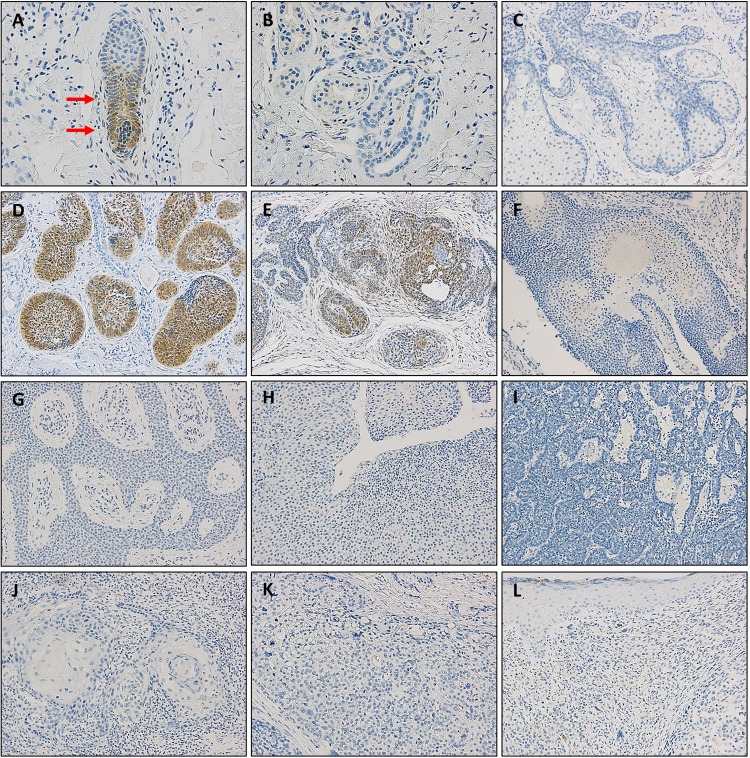
Fig 3. Immunohistochemical staining for GLI1 in normal skin and various skin tumors.
Representative images of GLI expression in hair follicle (A), sweat gland (B), sebaceous gland (C), basal cell carcinoma (D), trichoepithelioma (E), pilomatricoma (F), eccrine poroma (G), hidradenoma (H), spiradenoma (I), squamous cell carcinoma (J), sebaceous carcinoma (K), and malignant melanoma (L). A, B: ×400 magnification, C-L: ×200 magnification.

