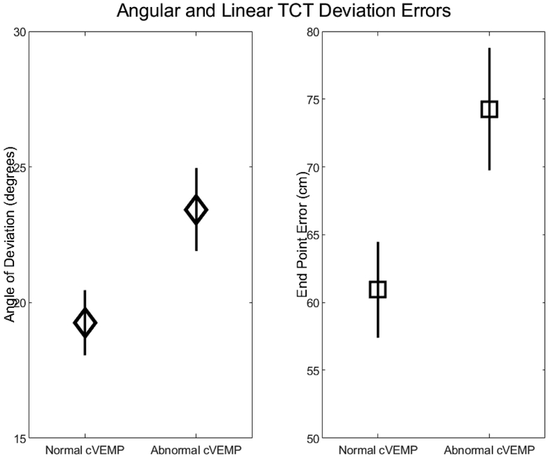
Figure 2.
Angular deviation and end point errors increase when saccular function is abnormal (unilaterally or bilaterally absent) compared to normal (bilaterally present). Diamonds and squares represent the marginal means for angular deviation (degrees) and end point error (cm) respectively, controlling for age and sex. Error bars represent standard errors.

