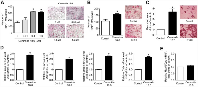
Figure 3.
Ceramide 18:0 stimulates osteoclast differentiation. (A) Primary mouse BMMs were incubated with 30 ng/mL M-CSF and 100 ng/mL RANKL in the absence or presence of the indicated concentration of C18:0 for four days. After staining cells with TRAP, the number of TRAP-positive multinucleated cells (MNCs) (≥3 nuclei/cell) was determined to assess osteoclast differentiation. (B) Mouse BMMs were cocultured with primary calvaria osteoblasts for 10 days in medium containing 10−8 M 1α,25-OH(2) D3 and 10−6 M prostaglandin E2 without or with 0.1 μM C18:0. (C) Mouse BMMs were cultured with 30 ng/mL M-CSF and 100 ng/mL RANKL on dentine discs in the absence or presence of 0.1 μM C18:0 for 10 days. Resorption pits were visualized by staining with hematoxylin. (D) qRT-PCR expression analysis of osteoclast differentiation markers in mouse BMMs exposed to 30 ng/mL M-CSF and 100 ng/mL RANKL in the absence or presence of 0.1 μM C18:0 for 4 days. (E) qRT-PCR analysis to determine relative Rankl and Opg expression in mouse calvaria osteoblasts exposed to 50 μg/mL ascorbic acid and 10 mM β-glycerophosphate in the absence or presence of 0.1 μM C18:0 for 7 days. Scale bars: 500 μm for (A–C). Data are presented as mean ± SEM. *P < 0.05 vs. untreated control using the Mann-Whitney U test or ANOVA followed by Tukey’s posthoc analysis.

