Figure 4.
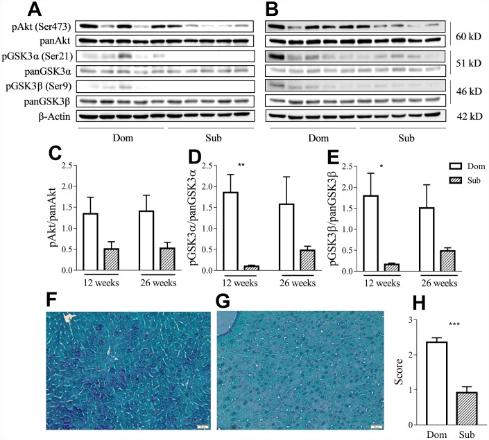
Sub mice exhibited decreased Akt and GSK3 phosphorylation and glycogen content in liver. Western-blot analysis of phospho- and pan-Akt and GSK3 levels in the liver of Dom and Sub mice at the age of 12 (A) and 26 weeks (B). (C) Phosphorylation level of Akt (Ser473) in Dom and Sub mice at the age of 12 and 26 weeks (n= 5 for each group). (D) The phosphorylation level of GSK3α (Ser21) in Sub mice was significantly decreased at the age of 12 weeks (Student unpaired two-tailed t-test, t=4.091, p< 0.01; n=5 for each group). (E) The phosphorylation level of GSK3β (Ser9) in Sub mice was significantly decreased at the age of 12 weeks (Student unpaired two-tailed t-test, t=3.012, p<0.05; n= 5 for each group). The amount of glycogen in the liver of Dom (F) and Sub (G) mice, PAS staining, scale bar - 20μm. (H) PAS staining revealed reduced level of glycogen in liver of Sub mice (Mann-Whitney test, p<0,001). The staining intensity of PAS staining was scored as 0 (0-25%), 1 (26-50%), 2 (51-75%), or 3 (76-100%) according to the percentage of positively stained cells. * - p<0.05, ** - p<0.01, *** - p<0.001. Error bars indicate SEM.

