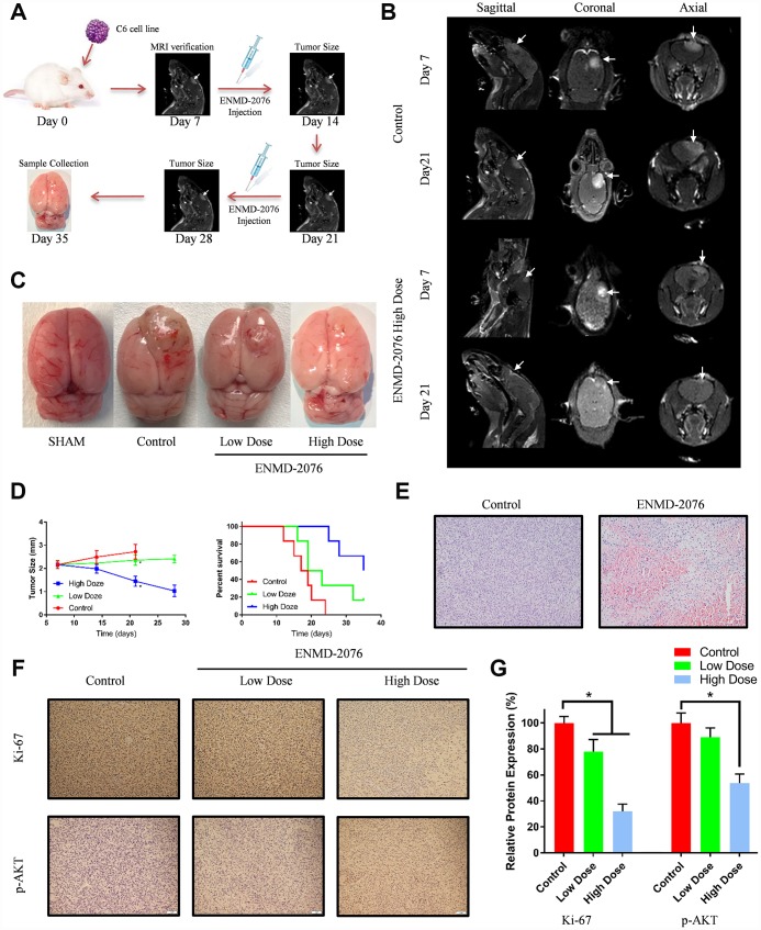
Figure 8.
(A) The overall framework of animal experiment. C6 cells were intracranial injected on day 0. Magnetic resonance imaging (MRI) was used to evaluate the tumor growth on day 7. (B) MRI examination with different planes (sagittal, coronal, axial). (C) The representative samples showing the tumor volumes in different groups. (D) Tumor size and percent survival curves in different groups. (E) Histologic features of control group and ENMD-2076 treated group. (F–G) Immunohistochemistry analysis of ki-67, p-AKT in the different groups.

