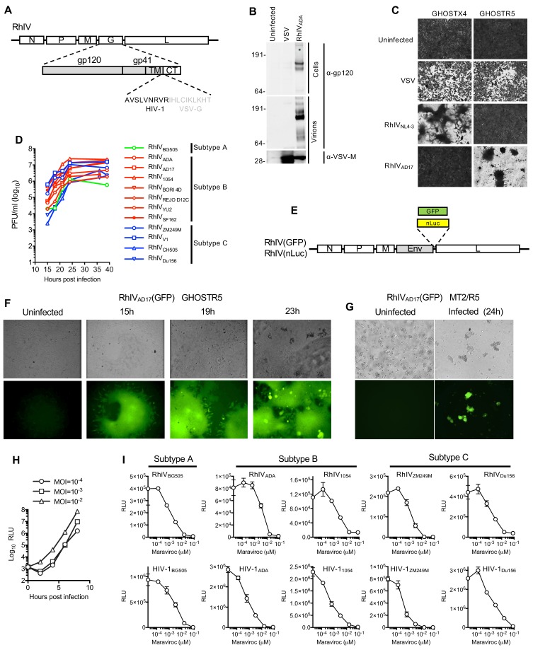
Figure 1. Characterization and in vitro replication properties of RhIV strains.
(A) Schematic representation of RhIV genomes in which VSV-G ectodomain and transmembrane sequences are replaced with HIV-1 Env counterparts. (B) Western blot analysis of HIV-1 gp160/120 and VSV-M protein levels in RhIV infected cells and extracellular virions. (C) Monolayers of GHOST-X4 or GHOST-R5 cells stained with crystal violet 24 hr after infection with VSV or RhIV strains. (D) Yield of various RhIV strains in plaque forming units/ml (PFU/ml) during replication in 293 T/CD4/CCR5 cells. (E) Schematic representation of RhIV genomes in which a GFP or nanoluciferase (nLuc) reporter is included. (F,G) Micrographs of GHOST-R5 (F) and MT4-R5 (G) at the indicated times after infection with RhIVAD17(GFP). (H) Luciferase expression in TZM-Bl cells over time following infection with RhIVAD17(nLuc) at the indicated MOIs. (I) Inhibition of RhIV(nLuc) and corresponsing HIV-1(nLuc) strains by the CCR5 inhibitor, Maraviroc.

