Figure 6.
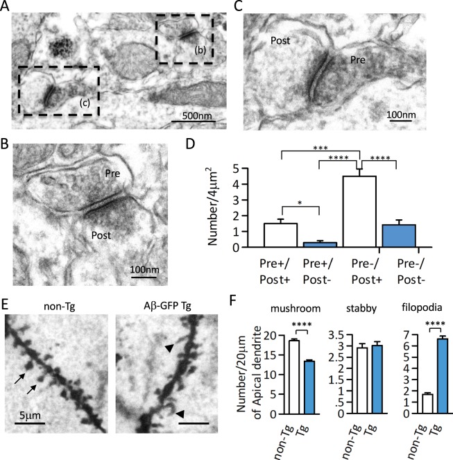
Synaptic expression of Aβ-GFP fusion protein and morphological changes in synapses in Aβ-GFP Tg mice. (A–C) Representative Immuno-EM image by anti-GFP antibody in Aβ-GFP Tg mice hippocampus at 3-month-old. The dotted rectangle in (A) are shown at higher magnification in (B) and (C). (D) Histograms representing the numbers of immunopositive synaptic structures per unit areas of stratum-radiatum in the hippocampal CA1 region. Aβ-GFP fusion protein was expressed mainly in the post synaptic sites (*p < 0.05, ***p < 0.0050, ****p < 0.0001 by one way ANOVA, n = 24 areas, data are presented as means ± SEM). (E) Golgi staining of apical dendrite from non-Tg and Aβ-GFP Tg mice hippocampal CA1 regions. Arrows showed spine and arrow heads showed filopodia. Compared to the non-Tg, Aβ-GFP Tg mice had significantly fewer spines and significantly more filopodia (****p < 0.0001 by Student’s t-test, n = 5 animals each, 20 μm length of dendrites from 24–27 areas each, data are presented as means ± SEM).

