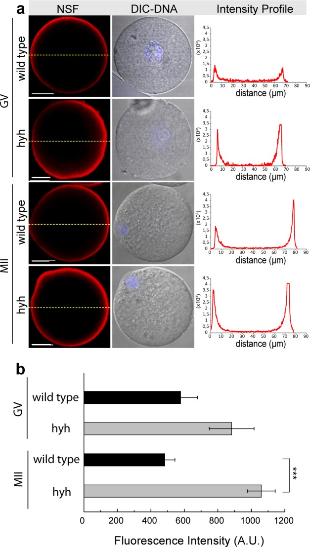
Figure 2.

NSF inmunodetection during meiotic maturation in wild type and hyh MII oocytes. (a) NSF was detected by indirect immunoflurescence in wild type and mutant homozygous (hyh) GV-intact oocytes (GV) and MII oocytes (MII). Red indicates positive staining for primary NSF antibody detected by a secondary antibody conjugated to Alexa Fluor 594; blue indicates DNA, labeled with Hoechst 3342; DIC shows differential interference contrast (DIC) images. Right column in each panel shows the fluorescence intensity profiles for NSF. Fluorescence intensities were measured along equatorial dashed yellow lines traced in each oocyte. The intensity of NSF is indicated by red lines. Scale bar: 20 μm. (b) Histogram showing fluorescence intensity quantification (A.U.: arbitrary units) for wild type GV and MII oocytes compared to hyh ones. ***p ≤ 0.001 (Student’s t-test).
