
Figure 5.
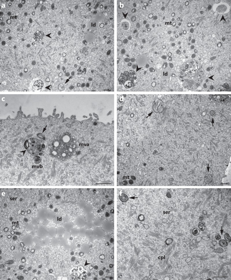
Cytoplasmic degradative structures in hyh oocytes. Representative electronmicrographs of MII oocytes from mutant homozygous (hyh) mice showing different degradative structures such as autophagic-like vesicles in different stages of maturation (arrowheads; a–c,e) and dense lamellar bodies (arrows; a,c,d,f). More immature autophagic-like vesicles show a double membrane separated by a wider lumen and its main contents appear to be recognizable cytoplasmic organelles. Meanwhile more mature autophagic-like vesicles are delimited by a single membrane containing membranous material of unrecognizable origin. Note in (c) that a multivesicular body (mvb) and a multilamellar body (arrow) are in close association with an autophagic-like vesicle (arrow head). Mt: mitochondria; ld: lipid droplet; mvb: multivesicular body; mva: multivesicular aggregate; cpl: cytoplasmic lattice; ser: smooth endoplasmic reticulum. Scale bar: 1 μm.