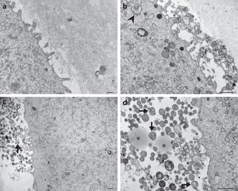
Figure 6.

Ultrastructure of perivitelline space in wild type and hyh MII oocytes. Representative transmission electron microscopy images of perivitelline spaces from wild type (a) and mutant homozygous (hyh) oocytes (b–d). (a) Perivitelline space from wild type MII oocytes is free of extracellular structures, only oolema microvilli are observed. (b–d) Numerous residual bodies are observed in the perivitelline space of mutant homozygous (hyh) MII oocytes, many of them are membrane-delimited. Additionally, remnants of cytoplasmic components as lipid droplets (asterisk) and dense lamellar bodies (arrow) are observed under higher magnification (d). Note phagophore-like structure surrounding a membrane bound vesicle (b, arrowhead). Scale bar: 1 μm.
