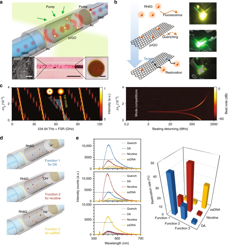
Fig. 1. Conceptual design and functionalization of the prGO inner-deposited fiber sensor.
a Schematic architecture of the device. Two reflectors are collimated in a silica capillary (outer diameter 3 mm, inner diameter 125 μm, and length 10 mm) with the prGO deposited inside. Rhodamine 6G (Rh6G) works as the optical-gain media. Inset from left-to-right shows the scanned electron micrograph of the prGO, the microscopic pictures of the resonator, and the fiber end with Au coverage. Inset scale bars from left-to-right are, respectively, 2, 500, and 50 μm. b FRET sensing process. Fluorescence quenches when Rh6Gs (orange dots) attached on the prGO intracavity first, afterwards the targets bind on the prGO, enabling the Rh6G fluorenscent restoration. Here we also show the pictures during this process from top to bottom, with fluorescent color in yellow. c In the FP resonator, multiple longitude modes belong to varied FSRs. The simulated FSRs of the 1st-order transverse mode and the 2nd-order transverse mode are shown, with the resulting frequency shift beat note. Here the thickness of prGO is assumed at 50 nm. d Functionalization of the prGO. By linking H+ (pH = 2), OH− (pH = 8), and Na+ (pH = 7) with the prGO via chemical bonding, FRET in the sensors shows high selectivity detection of dopamine (DA), nicotine, and ssDNA molecules, respectively. e Measured results for the sensor-target pairs. Specific pair has fluorescent restoration intensity higher than the others, thereby enabling electronic signal counts over the noise-limited threshold of ≈4000 intensity counts (measurement acquisition time, 1 s).

