Fig. 1.
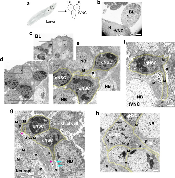
Drosophila larval brain qNSC protrusions contain clustered mitochondria as observed by TEM. a Drosophila larval brain. Larva, left; brain, right (schematic diagrams). Neuropil, gray regions in the brain lobe (BL) and thoracic ventral nerve cord (tVNC). b–h TEM images, amino-acid-depleted conditions. b Larval brain, low magnification. N, neuropil. c BL region (inset, gray box) at higher magnification in d and e, showing qNSCs (e, yellow overlays). f tVNC with mitochondria (M) in protrusions (P). g qNSC protrusions form junctions with the neuropil (magenta arrowheads); small mitochondria (cyan arrows) in the neuropil near a qNSC protrusion end. Putative glial cell (upper right) with an irregular cell body. Abn M, abnormal mitochondria (see Supplementary Fig. 1). h Clustered mitochondria in a qNSC protrusion neck (center). NB, neuroblast; Nu, nucleolus. Bars: 10 µm (b); 2 µm (c); 500 nm (d–h).

