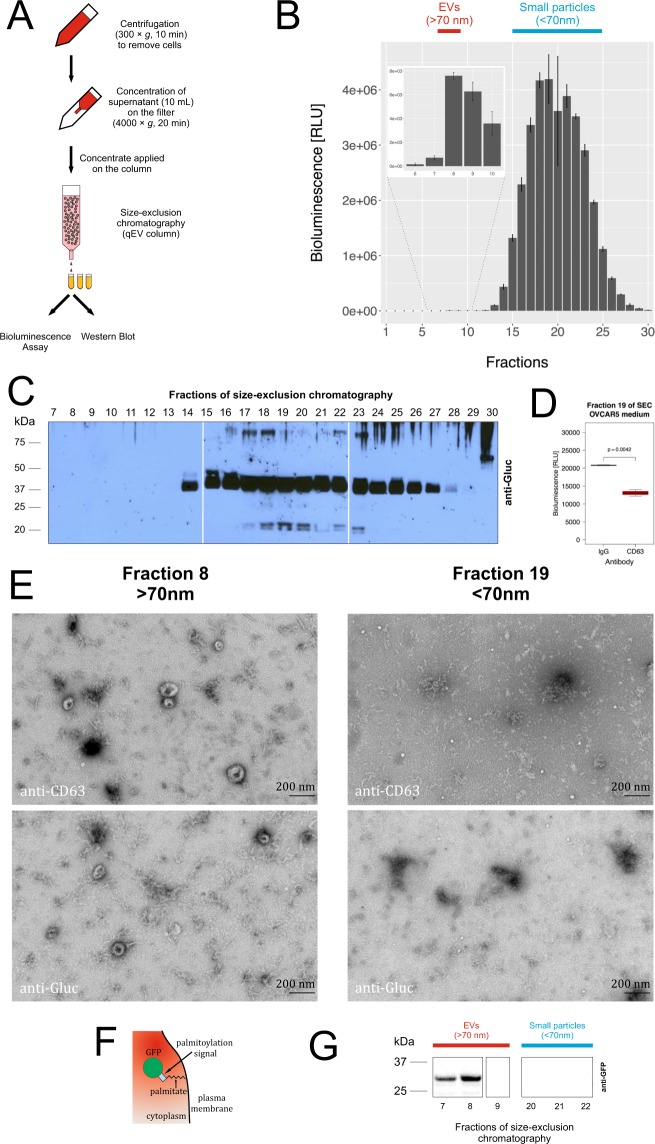
Figure 4.
mbGluc Is Mainly Present in the Small Particle – Fractions by Size Exclusion Chromatography (SEC). (A) Schematic of procedure to fractionate conditioned medium by SEC. (B) mbGluc activity in SEC fractions (qEV column) after fractionation of OVCAR5 conditioned medium (Procedure in (A); (representative of 2 experiments, each fraction measured in 3 technical replicates). The insert represents fractions 6–10 with a modified scale on Y axis that ranges from 0 to 8,000 RLU. (C) Western blot analysis of mbGluc protein following fractionation of OVCAR5-mbGluc+ medium by SEC (representative of 2 experiments). The main mbGluc band was at 40 kDa, similarly to that observed in the top fraction after sucrose step-gradient fractionation of the conditioned medium (compare to Fig. 2C). Two other less intense bands are at 20 kDa (which is close to the approximate size of free non-membrane bound Gluc without the biotin acceptor domain – 19.9 kDa) and 80 kDa (twice the size of the dominant 40 kDa band). The white bar divides lanes from separate membranes with samples from the same experiment processed according to the same procedure. Full picture of the gel is presented in Fig. S5E. (D) mbGluc activity in structures captured by anti-CD63 or IgG (unspecific binding) antibody in fraction No 19 which is representative of small particles fractions (fractionation of OVCAR5 medium in qEV column). (E) Transmission electron microscopy (TEM) of fraction 8 and 19 of SEC (qEV column). Material from the fractions was directly applied on the grid and immunolabeled with either anti-CD63 or anti-Gluc antibodies, scale bar: 200 nm. (F) Schematic diagram of cell membrane and EV labeling with palmGFP. (G) Western blot analysis of palmGFP protein following fractionation of OVCAR5-palmGFP + medium by SEC in fractions representative of EVs (7–9) and particles smaller than 70 nm (20–22). The white bar divides lanes from separate membranes with samples from the same experiment processed according to the same procedure. Full picture of the gel is presented in Fig. S5F.

