Figure 11.
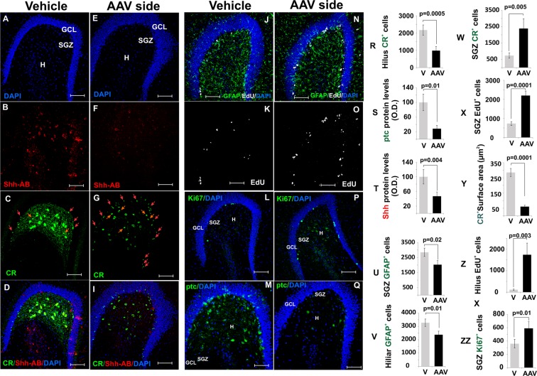
Effects of the Shh ablation on calretinin+ cells and proliferative markers. (20X, bar 100 um). (A–D) The vehicle-injected hippocampus show normal expression of Shh and CR proteins as detected by the antibodies (20X, bar 100 µm). (E–I) The AAV-injected hippocampus shows a substantial decrease of Shh levels and a striking reduction in size and numbers of CR+ cells. (20X, bar 100 µm). Also note the increase in small CR+ cells in the hilus (red arrows) and toward the hilus (yellow arrows) (see also T,W). (J–K) The vehicle-injected hippocampus shows normal expression of GFAP (glial cells) and EdU (20X, bar 100 µm). (N,O) The AAV-injected DG shows increased expression of EdU and a slight decrease in GFAP signaling (20X, bar 100 µm). (L,P) Ki67 also was found to be elevated in the AVV injected hilus (20X, bar 100 µm). (M,Q) Ptc protein expression decreased significantly followed the virus transfection (20X, bar 100 µm). (R–ZZ) Quantification of cells number and protein levels identify in the pictures above. The p scores correspond to unpaired (two tailed) t-test (n/group = 6). Abbr: GCL, granular cell layer; SGZ subgranular zone; and H, hilus.

