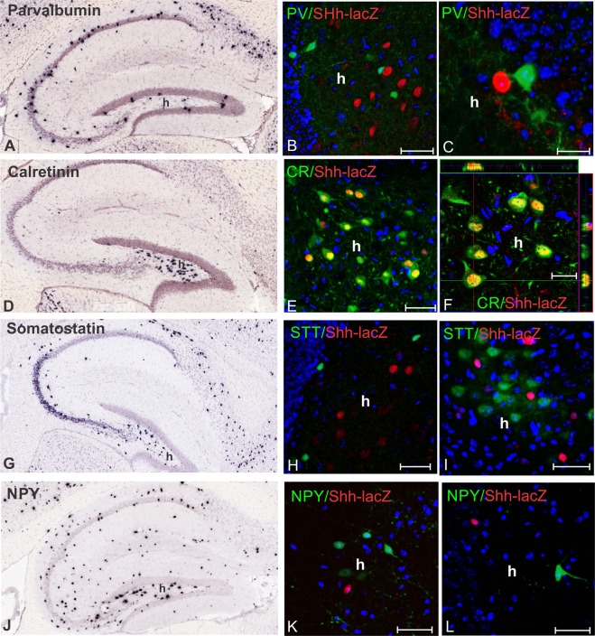
Figure 2.
GABAergic neurons expressing Shh-nlacZ-mRNA are calretinin immunoreactive (for quantification see Table 1). (A) In situ hybridization in a control mouse showing parvalbumin expression pattern in the hippocampus; (B,C) Antibody staining for parvalbumin (PV) and Shh-nlacZ in the hilus. PV and Shh-nlcZ, show no colocalization (B, 25X, bar 50 µm; C, 63X, 20 µm). (D) In situ hybridization showing calretinin expression pattern in the hippocampus. (E,F) Immunostaining for calretinin and Shh-nlacZ in the hilus showing colocalization (E, 25, bar 50 µm; F, 63X, 20 µm). (G) In situ hybridization showing somatostatin expression pattern in the hippocampus. (H,I) Immunostaining for somatostatin and Shh-nlacZ in the hilus showing no colocalization (25X, bars 50 µm). (J) In situ hybridization showing NPY expression pattern in the hippocampus. (K,L) Immunostaining for NPY and Shh-nlacZ (see Table 1) in the hilus shows no colocalization. (K, 25X, bars 50 µm). In situ hybridization images were obtained from Allen Mouse Brain Atlas (http://mouse.brain-map.org)21. Abbr: h, hilus.

