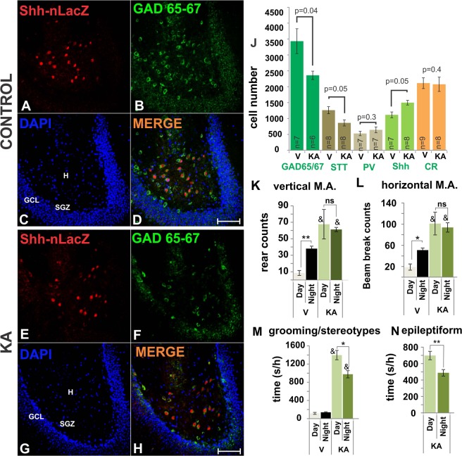
Figure 4.
KA administration induces loss of large number of GABA cells but GABAergic Shh-nlacZ+ neurons seem unaffected. (A–D) Shh-nlacZ+ GAD 65/67+ neurons in the DG of control animals showing co-localization (25X, bar 100 µm). Note the full complement of cells expressing Shh-lacZ in the hilus (red channel). (E–H) The number of Shh-nlacZ+ neurons did not decrease while the number of GAD 65/67+ neurons appears to diminish at 2 weeks following KA injections (25X, bar 100 µm). (J) Cells expressing Shh-nlacZ were up-regulated, while the overall expression of GAD 65/67 decreased. GABAergic subtypes included PV, parvalbumin; SST, somatostatin; CR, calretinin and Shh, sonic hedgehog. Examples of these staining are provided in Fig. 2. (p values correspond to Unpaired Student’s t-Test). (K–N) Behavior was analyzed on the second week after injection, scores correspond to average counts/h or seconds/h of a given behavior for 7 days, 4 h/day(12–4 pm) and 4 h/night (12–4am). *p < 0.01, **p < 0.001, night vs. day; & p < 0.001 KA vs. vehicle group. Epileptiform activity was always equal or lower than stage 3 (mouse forelimb clonus without rearing) (n = 6/group). Abbr: GCL, granular cell layer; SGZ subgranular zone; H, hilus; M.A., motor activity.

