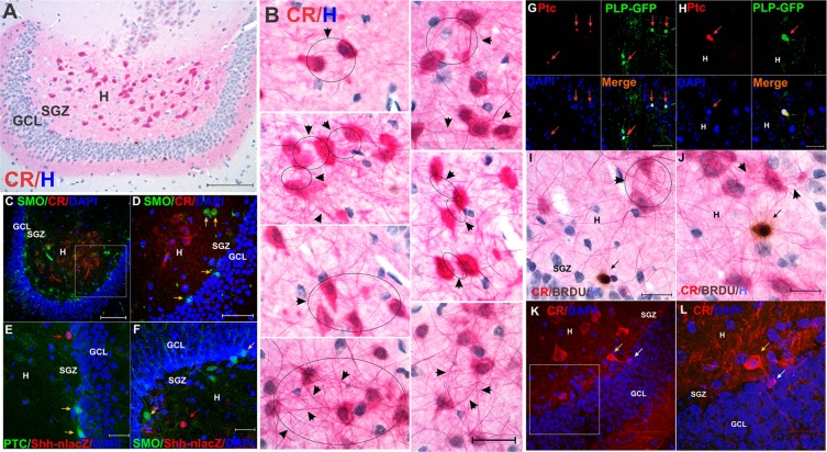
Figure 7.
Pattern of reciprocal innervation of ventral DG CR+ cells and expression of Shh receptors. (For quantification see Table 1). (A) Panoramic view showing abundant numbers or large CR+ neurons in the ventral DG (10X, bar 100 µm). (B) Detailed view of CR cells and their axonal/dendritic processes, showing profuse reciprocal connectivity (arrow-circles and arrowheads), which is a typical feature of CR (60X, bar 50 µm). (C) Expression of Smo and CR is segregated in the hilus. Most CR+ cells are located in the central hilus, while Smo+ cells are located in the peripheral SGZ (25X, bar 100 µm). (D) Close-up of C showing Smo positive cells (yellow arrows) in the SGZ (25X, bar 50 µm). (E) Shh-nlacZ (red arrow) and Ptc-1 (yellow arrow) do not co-express in the DG (for quantification see Table 1) suggesting separated cellular sources (63X, 25 µm). (F) Shh-nlacZ (red arrow) and Smo (yellow arrow) do not co-express in the DG (for quantification see Table 1) suggesting independent cellular sources (63X, 25 µm). (G) Ptc-1 is expressed by olidodendrocytes (PLP/Shh-nlacZ, 92.1 ± 4.7%, n = 3 × 10 slices) that do not co-express Shh (see Fig. 1D) (40X, bar 50 µm). (H) Larger magnification as in G (63X, bar 20 µm). (I) Hilar CR+ projections innervating a BRDU+ progenitor (black arrow) in the SGZ. Immunohistochemistry was performed with chromogenic staining for CR (Vulcan Fast Red), BRDU (DAB) and hematoxylin (H) as counterstaining (60X, bar 20 µm) (arrowheads and circle also show reciprocal innervation). (J) Hilar CR+ projections innervating a BRDU+ progenitor (black arrow) located in the central hilus. Immunohistochemistry was performed with chromogenic staining as in I (60X, bar 20 µm). (arrowheads show reciprocal innervations as in I). (K) Example of a calretinin (CR+) neuron from the hilus (yellow arrow) innervating a SGZ CR+ neural progenitors (white arrow) (63X, bar 50 µm). This suggests that CR+ neurons from the hilus innervate CR+ SGZ newborn neurons. (L) Close-up of K showing innervation of a CR+ progenitor (white arrow) by a CR+ neuron (yellow arrow) (63X, bar 20 µm). Abbr: DG dentate gyrus; GCL, granular cell layer; SGZ subgranular zone.

