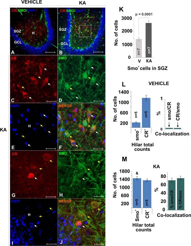
Figure 8.
Smo is expressed by hilar CR+ cells following KA treatment. (A,B) The Shh downstream molecule Smo was up-regulated in animals that received KA injections. In A, smo is preferentially detected in the SGZ but in B Smo and CR staining colocalized in many cells in the central hilus. (25X, scale bar 100 µm). (C–F) High magnification as in B (63X, bar 25 µm). Example of cells that express smo but do not express CR (white arrow) and CR+ cells that express both Smo and CR (yellow arrow). (G–J) More examples at high mag (63X, bar 25 µm) of cells that expresses smo but does not express CR (white arrow) and cells that express both Smo and CR (yellow arrow). Note that the small CR+ cell showing high intensity for CR and very weak staining for Smo is an SGZ-immature neuron. (K) Smo was significantly up-regulated in the SGZ in animals receiving an epileptogenic dose of KA. (L,M) Both Smo-CR and CR-Smo co-localization increased from near 0% to about 70% in animals injected with KA. Quantification of cells in the hilus across the ventral hippocampus in vehicle treated mice show that Smo expression was relatively small (as in A, restricted to the SGZ). Smo and CR colocalization was not observed. The number of cells expressing Smo was significantly greater in KA treated mice (M) as compare to Vehicle (L)(&, KA vs. Control, p = 0.00001, student T-test), while CR+ cells number was unchanged. The co-localization of the two markers was higher in the hilus of KA treated mice (CR/Smo = 70%, Smo/CR = 70%). Abbr: GCL, granular cell layer; SGZ subgranular zone; and h, hilus.

