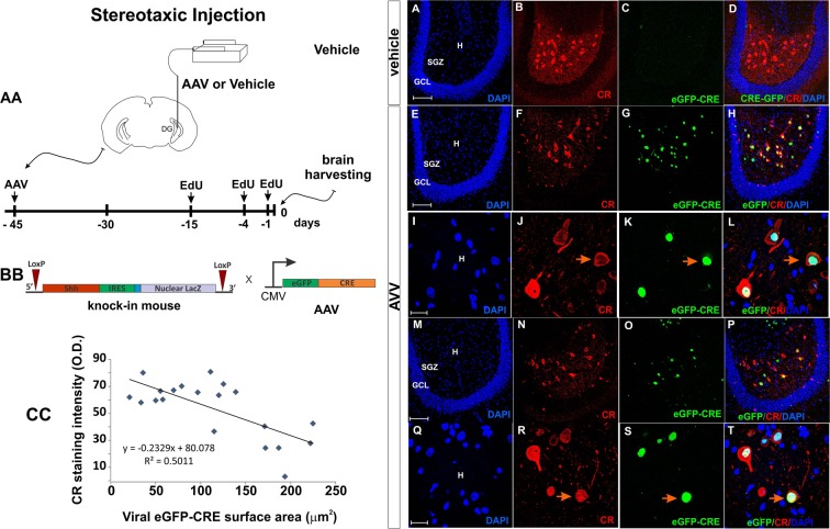
Figure 9.
Ablation of Shh using an AAV virus. (AA) Experimental design. The e-GFP-CRE AAV virus or vehicle was injected into the hilar region on one hippocampus (left side). Histological observations were made 45 days after the injections. EdU was administered 15, 4 and 1 days before euthanasia (See methods). (BB) Constructs of the Shh-nlacZ Knock-in and in the AAV vector. The expression of the virus in target cells will induce deletion of the Shh gene. (CC) Plot correlating the degree of viral infection as measured by the surface area of nuclear eGFP signal with CR staining intensity shows that cells that have greater infection levels express less CR. (A–H) Low magnification comparison between vehicle side and AAV injected side, 45 days after the injections of the AAV virus in the DG of Shh-nlacZ mice. The expression of the virus can be seen in G and how the virus targeted the CR+ neurons can be seen in H (20X, bar 100 µm). (I–L) High magnification images showing that the infection leads to reduced CR expression and cell loss (100X, bar 20 µm). (For quantification of CR cells see Fig. 11R). (M–P) Other examples at low mag as in E-H (20X, bar 100 µm). (Q–T) Other examples at high mag as in I-L (100X, bar 20 µm). Abbr: GCL, granular cell layer; SGZ subgranular zone; and H, hilus.

