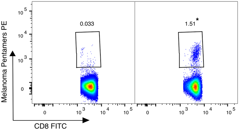
Fig. 1.
Patient 16 MT cultured from tumor-infiltrated lymph node (TILN) contains a melanoma associated antigen (MAA)-specific population. WT (left) and MT (right) TILN were incubated with HLA-A*0201 pentamers and co-stained for CD3 and CD8. Numbers in plots represent the frequency of MAA pentamer+ cells of total CD3+ CD8+ T cells. * frequency of MAA pentamer+ in MT was significantly greater than in WT (p < 0.001), modified Proschan’s method (Materials and Methods).

