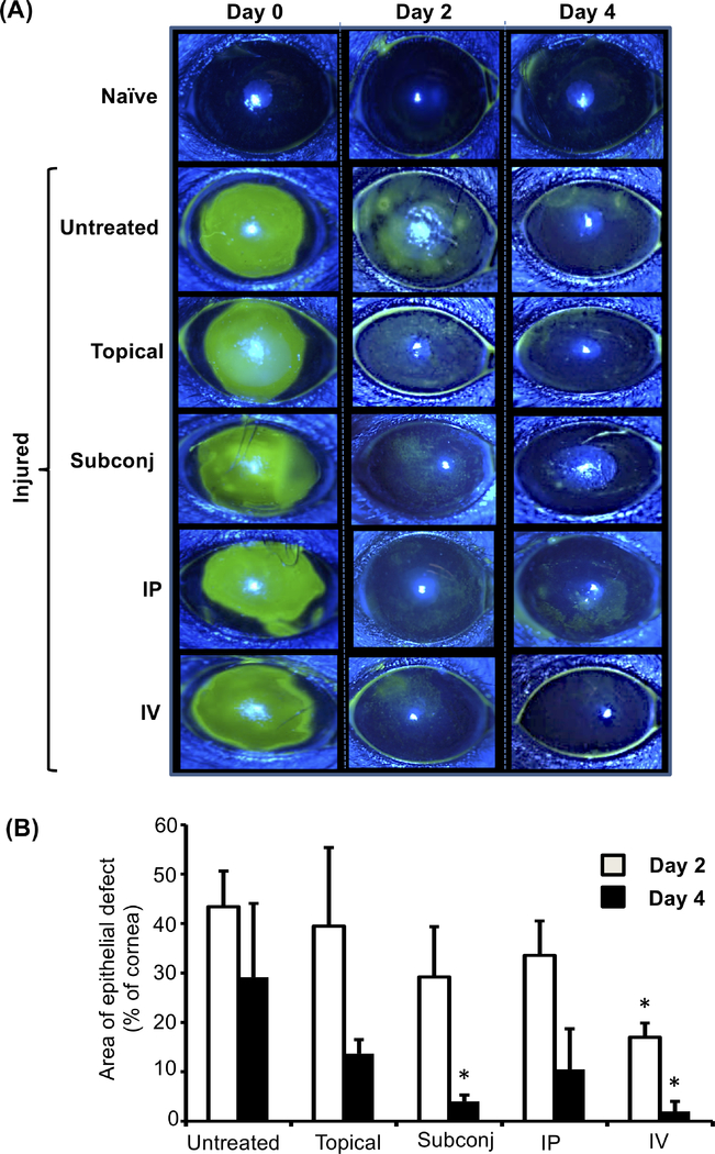
Figure 5. Subconjunctival or intravenous administration of MSCs accelerates corneal reepithelialization.
Corneal fluorescein staining of naïve and injured eyes was performed, and epithelial defects were evaluated by slit lamp biomicroscopy with cobalt blue light. (A) Representative images of fluorescein-stained corneas at days 0, 2, and 4 post-injury. The green areas represent epithelial defects. (B) Bar graph showing area of epithelial defect at days 2 and 4 post-injury (relative to day 0, set as 100%). Data from three independent experiments are shown, and each experiment consisted of 3–5 animals. The values are shown as mean ± SD. *p<0.05 as compared to untreated injured control.

