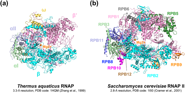
Figure 2. First high-resolution structures of RNAP.
A. First high resolution structure of a bacterial RNAP, T. aquaticus RNAP core at 3.3 Å resolution (Zhang et al., 1999). B. First high resolution structure of a eukaryotic RNAP, S. cerevisiae RNAP II core at 2.8 Å resolution (Cramer et al., 2001). All subunits are labeled on the figure and catalytic magnesium is drawn as an orange sphere in the center of the structures.

