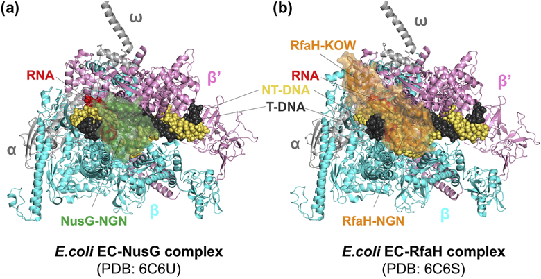
Figure 8. NusG- and RfaH-bound EC structures.
RNAP subunits and nucleic acids are labelled. A. E. coli NusG (colored in green) binds between β and β’ subunits, contacting β protrusion, β lobe, and β’ clamp helices. NusG KOW domain was disordered in the cryo-EM structure. B. E. coli RfaH (colored in orange) binds to the same site as NusG in panel A. In addition, RfaH recognizes a short-hairpin formed by ops sequence in the non-template DNA (not visible in the figure) and RfaH KOW domain binds flap tip of RNAP, covering upstream duplex DNA near the transcriptpion bubble.

