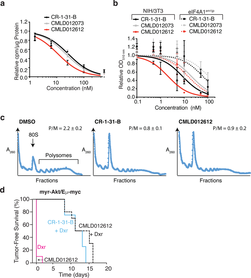
Figure 4. CMLD012612 inhibits tumor cell survival.
a. Inhibition of 35S-methioinine incorporation in HEK293 cells following 1 h compound exposure. n = 3 ± SEM. b. Cytotoxicity of CMLD012612 towards NIH/3T3 and eIF4A1em1JP cells following 4 day compound exposure. n = 3 ± SEM. c. CMLD012612 inhibits translation in vivo in the liver. Mice were injected with vehicle or CMLD012612 (0.5 mg/kg). Cytoplasmic extracts were prepared from livers 3 h later and resolved on 10%–50% sucrose gradients by centrifugation in an SW40 rotor at 150,000 × g for 2 h. Plotted are results of one representative experiment of two that showed similar results. The positions of 80S ribosomes and polysomes in the gradient are labeled, and the polysome/monosome (P/M) ratios indicated. d. CMLD012612 sensitizes myr-Akt/Eµ-Myc tumors to doxorubicin in vivo. Kaplan-Meier plot showing tumor-free survival of mice bearing myr-Akt/Eµ-Myc tumors following treatment with doxorubicin (Dox, red line; n = 10), CMLD012612 (solid black line; n = 10), CR-1–31-B + Dox (blue line; n = 4), or CMLD012612 + Dox (dashed black line; n = 10). p<0003 for CR-1–31-B+Dox versus Dox, and p<0.00001 for CMLD012612+Dox versus Dox.

