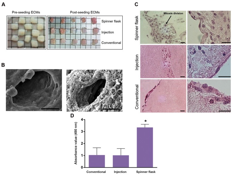
Fig 3.
Comparison of seeding protocols. A. Morphological, B. SEM, C. H&E staining, and D. MTS analyses. Comparison of recellularized human ovarian ECM with PMSCs through 3 seeding protocols (scale bars: 25 μm). ECM; Extra cellular matrix, MTS; 3-(4,5-dimethylthiazol-2-yl)-5-(3-carboxymethoxyphenyl)-2- (4-ulfophenyl)-2H-tetrazolium, SEM; Scanning electron microscopy and *; P<0.05.

