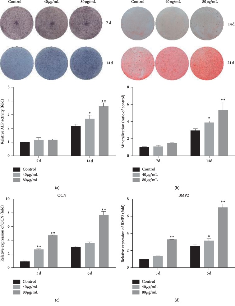
Figure 2.
The osteogenic effect of PCL on MC3T3-E1 cells. ALP staining (a) and Alizarin red staining (b) were carried out at 7 d and 14 d after treatment with PCL, respectively. Mineralized nodules were dissolved in cetylpyridine for relative quantification. Total RNA was extracted at 3 d and 6 d after treatment with PCL, and OCN (c) and BMP2 (d) expression was detected by RT-qPCR. ∗P < 0.05, ∗∗P < 0.01.

