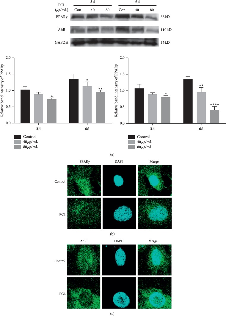
Figure 8.
Protein expression of PPARγ and AhR after treatment with PCL. (a) Total protein was extracted at 3 d and 6 d after treatment with PCL, and the relative expression levels of PPARγ and AhR were detected by western blotting. (b) Cell immunofluorescence staining was performed after treatment with PCL for 24 h; ∗P < 0.05, ∗∗P < 0.01, ∗∗∗∗P < 0.0001.

